
For those people that are a curious about the appearance of a normal colon, the following picture by picture account is offered for educational purposes.
The person shown in this "atlas of the colon" had a few polyps and a relatively easily photographed colon. The colonoscope was first pushed all the way through the colon. Pictures were taken as the colonoscope was withdrawn. When looking at these pictures any "white" areas are normal reflections from the light source.
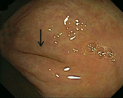
This first picture is of the opening of the appendix up close. The appendix is located in the furthest portion of the colon called the "cecum". This typically is the furthest point reached when screening the colon for cancer.
The next picture is taken a little further back. You can see the same slit like opening of the appendix on the right. The two large folds surrounding the appendix are called the "crow's foot". This is a normal landmark that further confirms that the end of the colon has been reached.
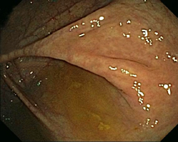
This is a picture of the cecum. The "crow's foot" is seen inside. The large, yellowish fold of tissue on the right is called the ileocecal valve. This is where the colon attaches to the small bowel.
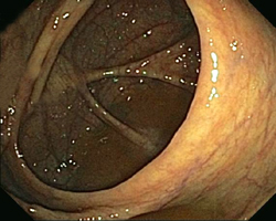
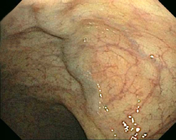
Moving backwards, the next portion of the colon is called the "ascending colon". You can see the cecum in the distance (compare to above picture). The large folds of the colon are called "haustral folds". These are normal. Notice the veins in the wall of the colon. This is healthy and normal as well.
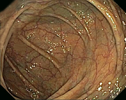
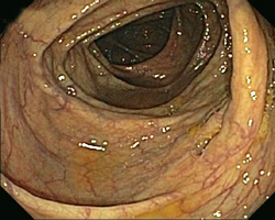
Moving further backwards, the first major turn happens where the liver is located. This is called the "hepatic flexure". The wall of the colon is slightly darker here at the top of the picture which is where the liver presses against the colon.
![]()
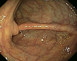
The next picture is of the "transverse colon". It is so named because it connects the right side of the colon to the left side, horizontally across the abdomen. It commonly has a triangle appearance as the picture on the left shows. The hepatic flexure is seen in the distance.
The second large turn of the colon occurs at the "splenic flexure". This is where the spleen is located in the abdomen. The transverse colon connects off the left side of the picture.
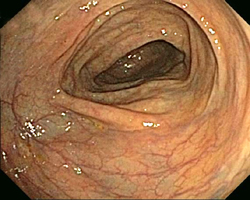
The next area of the colon is the descending colon". This is the portion of the colon which runs down the left side.
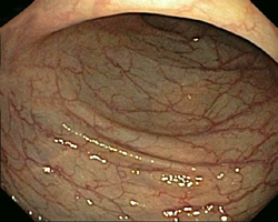
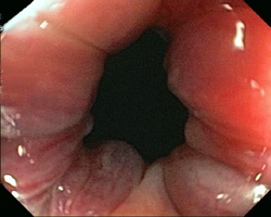
The last major portion of the colon is called the "sigmoid colon". This portion contains many turns as the picture on the right shows. It frequently causes abdominal cramping when an individual is constipated.
This is a picture of the rectum. Commonly large veins can be seen underneath the mucosa of its wall.
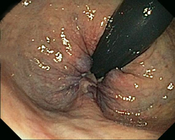
This is the rectum from the opposite angle. The black stripe is the colonoscope. The camera is pointed back on itself. The bulging areas at the base are hemorrhoids. The person here has moderate sized hemorrhoids.
This last picture is the anal canal. Here we can see the large, red hemorrhoids as seen above.
Next Chapter 5. Common conditions of the colon.